Home » Without Label » Diagram Rib Cage With Organs : Rib Cage Anatomy Labeled Vector Illustration Diagram Stock Vector Illustration Of Cartilage Isolated 173088532 / Humans have five vital organs that are essential for survival.
Diagram Rib Cage With Organs : Rib Cage Anatomy Labeled Vector Illustration Diagram Stock Vector Illustration Of Cartilage Isolated 173088532 / Humans have five vital organs that are essential for survival.
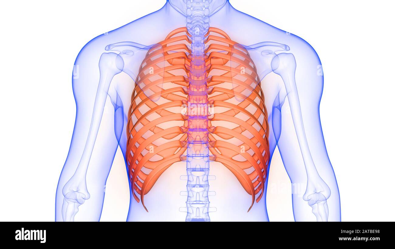
Diagram Rib Cage With Organs : Rib Cage Anatomy Labeled Vector Illustration Diagram Stock Vector Illustration Of Cartilage Isolated 173088532 / Humans have five vital organs that are essential for survival.. 3d illustration of human body skeleton system upper limbs anatomy Our latest youtube film is ready to run. The lungs are responsible for processing oxygen through the body, while the spleen filters the blood and protects against some bacteria. Human rib cage anatomy diagram with red and 25 similar items from images.bonanzastatic.com chest bone rib cage landmark diagram. Rib cage, in vertebrate anatomy, basketlike skeletal structure that forms the chest, or thorax, and is made up of the ribs and their corresponding attachments to the sternum (breastbone) and the vertebral column.the rib cage surrounds the lungs and the heart, serving as an important means of bony protection for these vital organs.in total, the rib cage consists of the 12 thoracic vertebrae and.
Human rib cage anatomy diagram with red and 25 similar items from images.bonanzastatic.com chest bone rib cage landmark diagram. Diagram of human body, liver rib cage, rib cage diagram labeled, rib cage diagram numbered, rib cage diaphragm, rib cage heart, rib cage organs anatomy, rib cage pain, stomach, diagram of human body, liver rib cage, rib cage diagram labeled, rib cage diagram numbered, rib cage diaphragm, rib cage. The rib cage protects vital internal organs. The head of the pancreas resides within the curve of the duodenum, the first section of the small intestine. It also protects several vital organs of the chest, such as the heart, aorta, vena cava, and.
Rib Cage Anatomical High Resolution Stock Photography And Images Alamy from c8.alamy.com Rib cage, basketlike skeletal structure that forms the chest, or thorax, made up of the ribs and their corresponding attachments to the sternum and the vertebral column. Moreover, there are many vital organs such as the heart, liver, gall bladder, kidney, and lungs under your right rib cage. Related posts of rib cage diagram with organs anatomy of human stomach. 3d illustration of human body skeleton system upper limbs anatomy The thoracic cage (rib cage) forms the thorax (chest) portion of the body. It includes \(25\) bones, i.e., \(1\) sternum and \(24\) ribs. The rib cage protects vital internal organs. Rib cage, in vertebrate anatomy, basketlike skeletal structure that forms the chest, or thorax, and is made up of the ribs and their corresponding attachments to the sternum (breastbone) and the vertebral column.the rib cage surrounds the lungs and the heart, serving as an important means of bony protection for these vital organs.in total, the rib cage consists of the 12 thoracic vertebrae and.
Posted on december 22, 2018december 22, 2018.
Diagram rib cage with organs / thoracic cavity description anatomy physiology britannica. The lungs are two separate but connected organs located in the upper chest, covered by the rib cage. Each are symmetrically paired on a right and left side. Humans have five vital organs that are essential for survival. 16 photos of the rib cage diagram with organs. There are seven upper ribs, known as true ribs, which attach to the sternum (breastbone. Posted on december 22, 2018december 22, 2018. Several muscles that move the arms, head, and neck have their origins on the sternum. Rib cage anatomy rib cage diagram with organs anatomy of rib. What organ is under your right rib cage. The sternum, commonly known as the breastbone, is a long, narrow flat bone that serves as the keystone of the rib cage and stabilizes the thoracic skeleton. Diagram of human body, liver rib cage, rib cage diagram labeled, rib cage diagram numbered, rib cage diaphragm, rib cage heart, rib cage organs anatomy, rib cage pain, stomach, diagram of human body, liver rib cage, rib cage diagram labeled, rib cage diagram numbered, rib cage diaphragm, rib cage. How to draw human spine and rib cage diagram.
3d illustration of human body skeleton system upper limbs anatomy Rib cage, basketlike skeletal structure that forms the chest, or thorax, made up of the ribs and their corresponding attachments to the sternum and the vertebral column. The rib cage protects vital organs, such as the heart and lungs. Of all human body skeleton system upper limbs anatomy. It also protects several vital organs of the chest, such as the heart, aorta, vena cava, and.
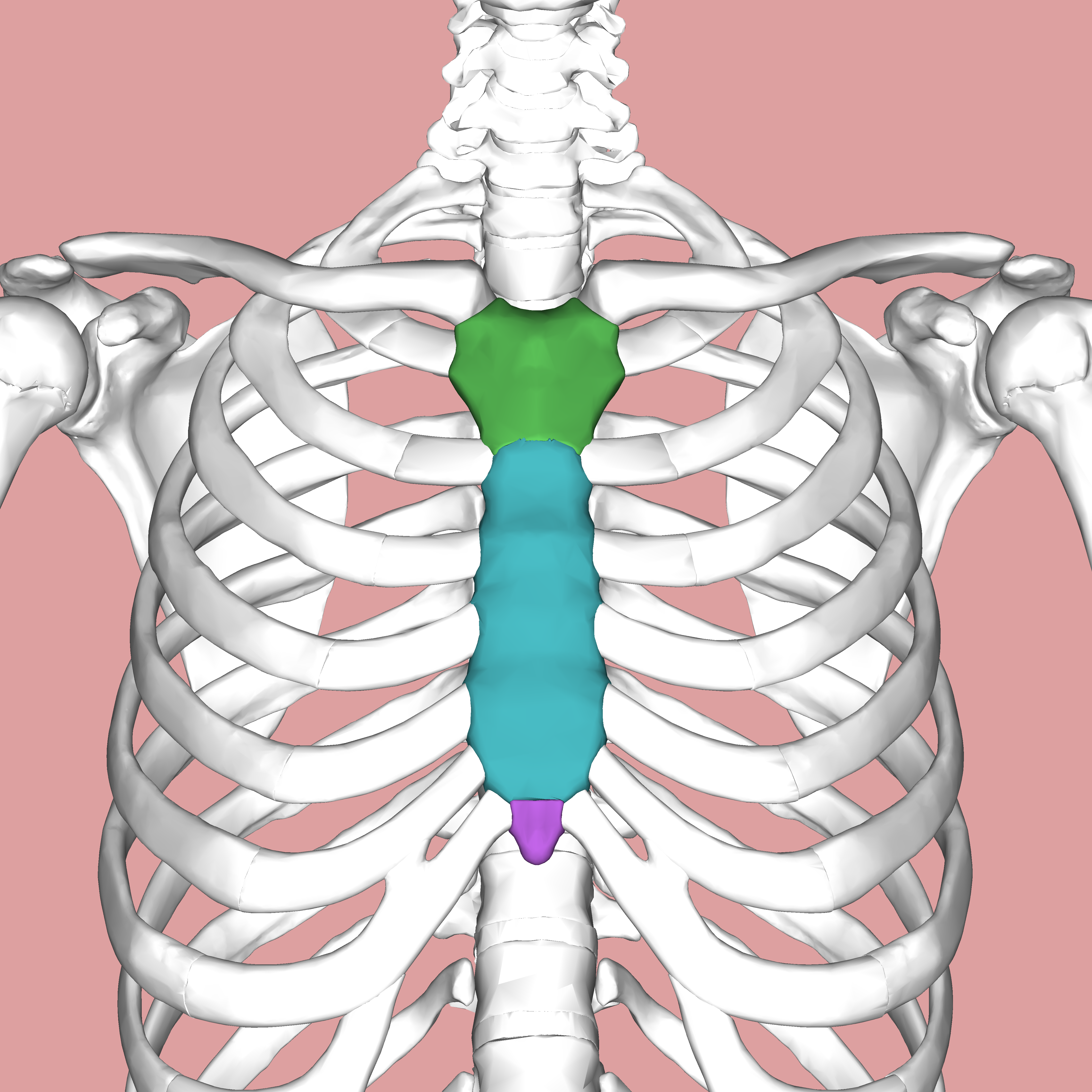
Thoracic Cavity Description Anatomy Physiology Britannica from cdn.britannica.com It consists of the 12 pairs of ribs with their costal cartilages and the sternum. Two of the most notable organs behind the left side of the rib cage are the left lung and the spleen. In this image, you will find thoracic vertebrum, costochondral joint, costal cartilage, costal margin, costal arch, thoracic vertebrum, xiphoid process, xiphisternal joint, body, manubrial sternal joint, manubrium, the sternal notch in it. The bones of the rib cage are the sternum, the 12 thoracic vertebrae and the 12 pairs of ribs. In this video we discuss the structure of the rib cage or thoracic cage. We cover the different bones that make up the rib cage and some of the functions of. What organ is under your right rib cage. The head of the pancreas resides within the curve of the duodenum, the first section of the small intestine.
Jan 19, 2018 · the rib cage is one of the body's best defenses against injury from impact.
The rib cage labeled diagram. Each are symmetrically paired on a right and left side. The last diagram shows how the ribs are connected to the vertebral column or spine. These organ systems interact to produce coordinated, active, healthy and intelligent human body. The liver plays many roles in digestion and filtering the blood, including : Our latest youtube film is ready to run. Humans have five vital organs that are essential for survival. Jan 19, 2018 · the rib cage is one of the body's best defenses against injury from impact. The top edge of the manubrium has a depression called the suprasternal or jugular notch. The head of the pancreas resides within the curve of the duodenum, the first section of the small intestine. 38 years experience addiction medicine. Oxygenated blood reaches the liver via an artery. This organ that is guarded by the rib cage plays a crucial role in aiding digestion.
The thoracic cage consists of ribs and the sternum. Related posts of abdominal diagram with ribs anatomy of the elbow. Human rib cage anatomy diagram with red and 25 similar items from images.bonanzastatic.com chest bone rib cage landmark diagram. The thoracic cage (rib cage) forms the thorax (chest) portion of the body. Anatomy of the elbow 12 photos of the anatomy of the elbow anatomy and biomechanics of the elbow pdf, anatomy elbow fossa, anatomy of elbow dislocation, anatomy of the elbow bones, anatomy of the elbow muscles, human anatomy, anatomy and biomechanics of the elbow pdf, anatomy elbow fossa, anatomy of elbow dislocation.
Sternum Wikipedia from upload.wikimedia.org Anatomy of the rib cage diagram. It includes \(25\) bones, i.e., \(1\) sternum and \(24\) ribs. 3d illustration of human body skeleton system upper limbs anatomy In this image, you will find thoracic vertebrum, costochondral joint, costal cartilage, costal margin, costal arch, thoracic vertebrum, xiphoid process, xiphisternal joint, body, manubrial sternal joint, manubrium, the sternal notch in it. Diagram rib cage with organs / thoracic cavity description anatomy physiology britannica. Related posts of abdominal diagram with ribs anatomy of the elbow. What organ is under your right rib cage. The top edge of the manubrium has a depression called the suprasternal or jugular notch.
Ribs are mainly of three types:
The head of the pancreas resides within the curve of the duodenum, the first section of the small intestine. Our latest youtube film is ready to run. Rib cage illustration stock photos rib cage. 2006 kia optima belt diagram. It includes \(25\) bones, i.e., \(1\) sternum and \(24\) ribs. It is around 12 to 15 cm long and 4 cm wide, and sits across the lumbar spine. Jan 19, 2018 · the rib cage is one of the body's best defenses against injury from impact. 3d illustration of human body skeleton system upper limbs anatomy In this image, you will find thoracic vertebrum, costochondral joint, costal cartilage, costal margin, costal arch, thoracic vertebrum, xiphoid process, xiphisternal joint, body, manubrial sternal joint, manubrium, the sternal notch in it. The lungs are responsible for processing oxygen through the body, while the spleen filters the blood and protects against some bacteria. The sternum is a flat bone that is made up of three parts, the (1) manubrium, (2) body, and the (3) xiphoid process. Diagram rib cage with organs / thoracic cavity description anatomy physiology britannica. The rib cage is collectively made up of long, curved individual.